General X-Ray
An X-ray is a painless, non-invasive procedure that creates images of a patient's internal structures to diagnose and treat illness or injury accurately. It's the most commonly used type of medical imaging.
During an exam, the X-ray machine sends a minute amount of radiation at the examination site. The radiation passes through the body and captures an image on a computer display. X-rays help doctors identify and treat a broad range of conditions, including:
- Broken bones
- Arthritis
- Joint injuries
- Pneumonia
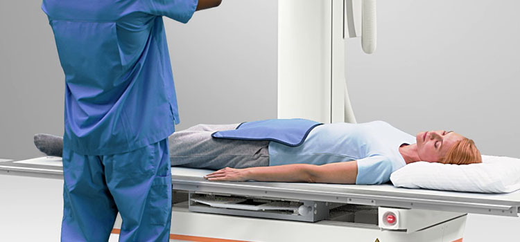
What should I expect when undergoing X-rays?
X-ray examinations are typically are quick and painless. The patient is exposed to as little radiation as possible while still generating acceptable images.
How do I prepare for an X-ray?
Arrive at least 30 minutes prior to the scheduled session to ensure that any necessary paperwork is completed. Remove all jewellery /items that may interfere with the procedure.
Make a radiographer aware of conditions/procedures you may have had, which are relevant prior to scanning, i.e.
- Pacemaker
- Pregnancy
- Implanted devices (i.e., chemo port /dialysis catheters)
- Artificial heart valves
- Aneurysm clips
- Cochlear implants
- Metallic implants and prosthesis
- Vascular stent or stent-graft.